
Fig. 2. MCH and hypocretin are not co-localized in the cat hypothalamus.
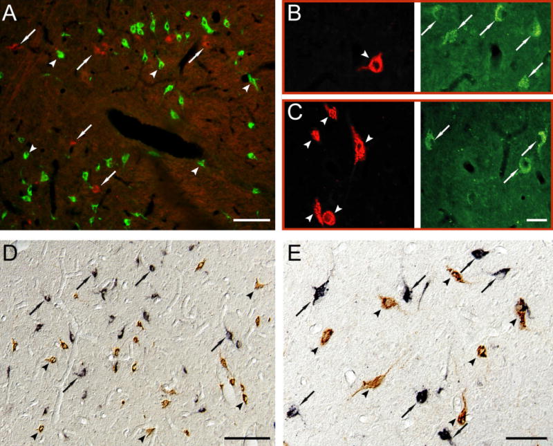
A. MCHergic (in green, examples in arrowheads) and hypocretinergic neurons (in red, examples in arrows) are intermingled in the postero-lateral hypothalamic area of the cat. The picture was obtained by superimposing photomicrographs that had been taken using green and red filters. B–C. Photomicrographs have been taken with two different filters in order to recognize MCHergic (left column, in red, arrowhead) and hypocretinergic neurons (right column, in green, arrows) in the postero-lateral hypothalamic area of the cat. The location of both neuropeptides is clearly different. These photomicrographs were taken from 20 μm-thick sections that were processed for immunoflourescence. Rhodamine and FITC were used as fluorescent agents. D and E. Photomicrographs exhibit MCHergic neurons (in brown, arrowheads) intermingled with hypocretinergic neurons (in black, arrows) in the postero-lateral hypothalamic area. The photomicrographs were obtained using Nomarski optics from 20 μm-thick sections, which were processed for immunohistochemistry utilizing the ABC-DAB method. Hypocretin immunostaining was enhanced by nickel. Calibration bars: A, 50 μm; B–C, 30 μm; D, 100 μm; E, 50 μm.