
Fig. 3. Location of MCHergic and hypocretinergic neurons in the hypothalamus of a representative cat.
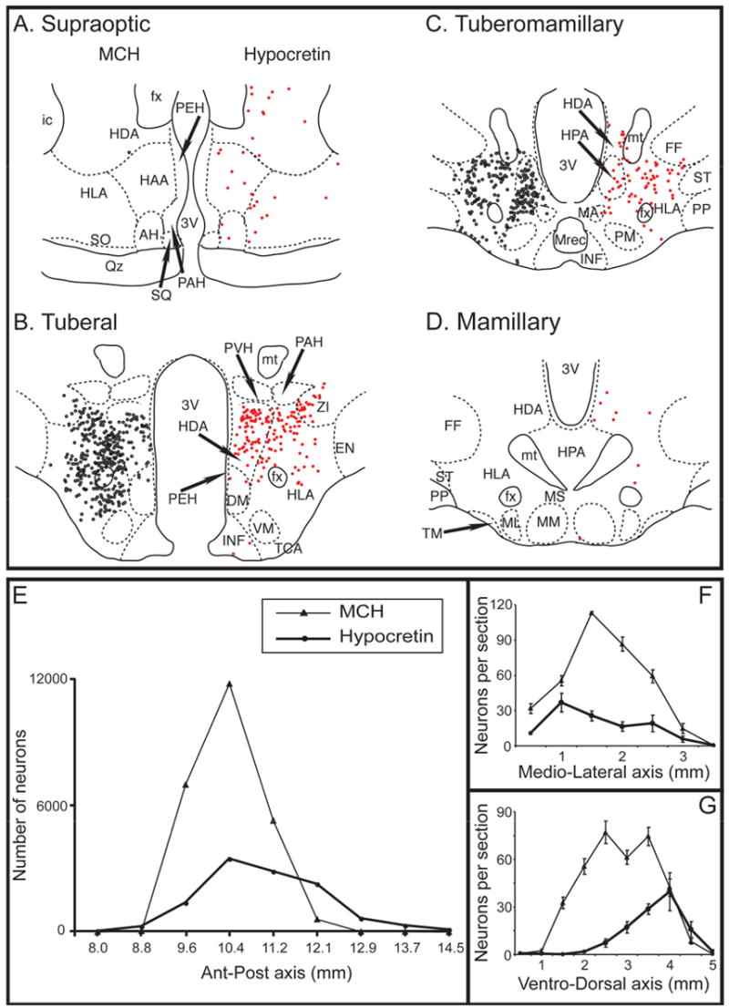
Camera lucida drawings of MCHergic (on the left, black circles) and hypocretinergic neuronal bodies (on the right, red circles) at different levels (A to D) of the hypothalamus are shown. The neurons are from the same hemi-hypothalamus (reflected in the figure). Both types of neurons were intermingled and were more concentrated at the tuberal level. There were no MCHergic neurons and very few hypocretinergic neurons at the preoptic level (not shown). Camera lucida drawings were obtained from adjacent sections, one was immunostained for MCH and the other was immunostained for Hcrt-2; these sections were counterstained with Pyronin-Y. The demarcation and nomenclature of cell groups in the cat hypothalamus are based on Berman and Jones, as well as Bleier's work (Berman and Jones, 1982, Bleier, 1961). E. Chart displaying the estimation of MCHergic (thin lines, triangles) and hypocretinergic neurons (thick lines, circles) in the antero-posterior axis of the whole hypothalamus (bilateral count). In F and G, the mean (± SE) number of neurons per hemisection in the medio-lateral and ventro-dorsal axes (analyzed at the tuberal level) is shown. In F, the horizontal axis is the distance from the medial surface of the hypothalamus to the lateral region. In G, the horizontal axis is the distance from the ventral surface of the hypothalamus to the dorsal region. AH, anterior hypothalamus; DM, dorsomedial nucleus; EN, entopeduncular nucleus; fx, fornix; FF, nucleus of the fields of Forel; HAA, anterior hypothalamic area; HDA, dorsal hypothalamic area; HLA, lateral hypothalamic area; HPA, posterior hypothalamic area; ic, internal capsule; INF, infundibular nucleus; MA, anterior mammillary nucleus; MM, medial mammillary nucleus; ML, lateral mammillary nucleus; M. Rec., mammillary recess of the third ventricle; MS, supramammillary nucleus; mt, mammillothalamic tract; PAH, paraventricular nucleus; PEH, periventricular complex; PM, premammillary nucleus; PP, pes pedunculi; PVH, parvocellular nucleus; Qz, optic chiasm; SQ, suprachiasmatic nucleus; SO, supraoptic nucleus; ST, subthalamic nucleus; TCA, area of the tuber cinereum; TM, tuberomammillary nucleus; VM, ventromedial nucleus; ZI, zona incerta; 3V, third ventricle.