
Fig. 4. Reciprocal axodendritic and axosomatic contacts between MCHergic and hypocretinergic neurons.
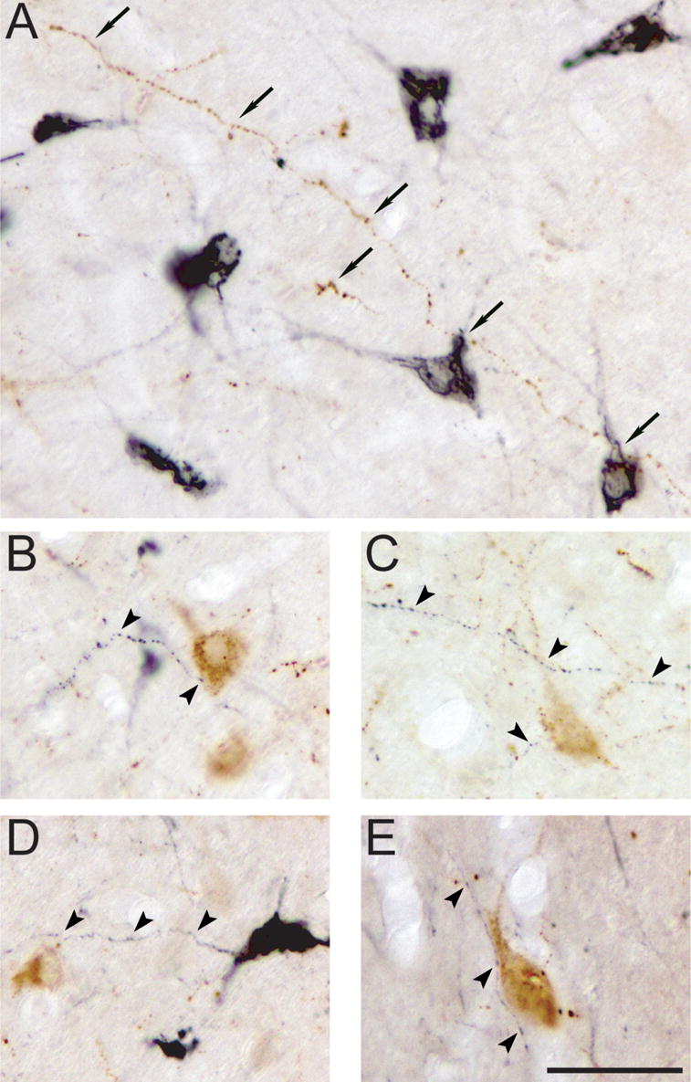
A. Apposition between a long hypocretinergic fiber (in brown, arrows) and MCHergic somata and primary dendrites (in black). B to E. MCHergic fibers (in black, arrowheads) were in close relationship with hypocretinergic somata and/or primary dendrites (in brown). All photomicrographs were taken from 20 μm-thick sections and processed with the ABC-DAB method. MCH immunostaining was enhanced by nickel. Calibration bar: 50 μm.