
Fig. 5. MCHergic neurons do not show Fos immunoreactivity during wakefulness or sleep.
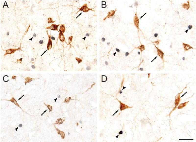
A–D. Immunoreactivity for Fos and MCH in sections of animals euthanized during A) active wakefulness with motor activity, B) AS induced by carbachol, C) active wakefulness without motor activity and D) quiet sleep. MCH-containing neurons exhibit brown immunoreactivity (examples are indicated by arrows); Fos immunoreactivity, which was restricted to nuclei, stained in black (examples are indicated by arrowheads). MCHergic neurons did not exhibit Fos immunoreactivity in any of the behavioral states that were examined. All photomicrographs were taken using Nomarski optics from 20 μm-thick sections and processed with the ABC-DAB method. Fos immunostaining was enhanced by nickel. Calibration bar: 40 μm.