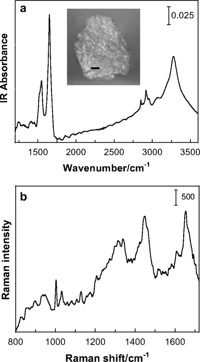
Fig. 1.

Vibrational microscopy of human corneocytes. a IR point mode spectrum (1,150–3,600 cm−1 region) acquired using a 40 μm2 aperture of a single corneocyte isolated from the third sequential tape strip applied to human forearm skin. The inset shows the optical image of the corneocyte (bar = 10 μm). b Raman spectrum (800–1,720 cm−1 region) of a similarly isolated corneocyte displaying bands characteristic of the lipid and protein (keratin) components
