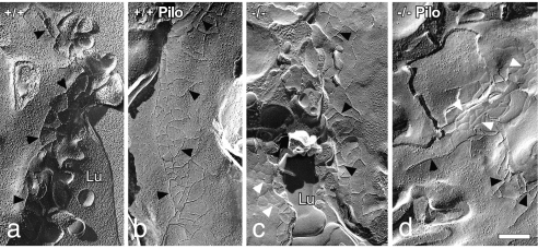
Fig. 4.
Freeze-fracture electron microscopy of parotid acinar cell tight junctions of male mice indicates an increase in strand number after pilocarpine stimulation. (a) AQP5+/+. (b) AQP5+/+, pilocarpine stimulated. (c) AQP5−/−. (d) AQP5−/−, pilocarpine stimulated. Black arrowheads indicate P-face junctional strands; white arrowheads indicate E-face grooves. Lu, lumen. (Scale bar = 0.25 μm.)

