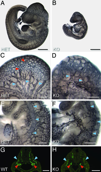
Fig. 2.
Defects in vascular remodeling in VE-PTPLz/Lz embryos. (A–F) Whole-mount PECAM-1-stained E9.5 embryos. VE-PTPLz/+ (A, C, and E) compared with a VE-PTPLz/Lz littermate (B, D, and F). (C and D) A closeup of the head vasculature shows the normal remodeling pattern of veins (blue arrows) and arteries (red arrows) in VE-PTPLz/+ embryos (C). In VE-PTPLz/Lz embryos, the venous plexus (blue arrows) undergoes only minor remodeling (D). (E and F) Closeup of the cardinal vein (blue arrows), which does not form correctly in VE-PTPLz/Lz embryos (F). (G and H) PECAM-1 immunostaining of E9.0 embryo cross sections at heart level. The dorsal aortas (red arrows) are small and collapsed in VE-PTPLz/Lz embryos (H) compared with WT (G), and the cardinal veins (blue arrows) are diffuse and not clearly delineated.

