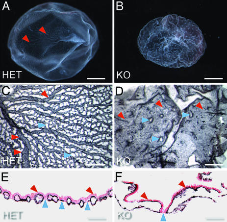
Fig. 3.
Vascular defects in yolk sacs of VE-PTPLz/Lz embryos. (A and B) Freshly dissected E9.5 VE-PTPLz/Lz yolk sacs (B) can be clearly distinguished from VE-PTPLz/+ littermates (A), because of a wrinkled appearance and a lack of visible vessels, which can be seen in the yolk sacs from VE-PTPLz/+ littermates (A, red arrows). (C and D) Whole-mount PECAM-1 staining at E9.5 confirms the lack of normal vessels in VE-PTPLz/Lz yolk sacs (D) compared with VE-PTPLz/+ (C). The endoderm and mesoderm layers in VE-PTPLz/Lz yolk sacs are barely connected (D, blue arrows), and the entire surface is lined with PECAM-1-positive ECs (red arrows). The VE-PTPLz/+ yolk sacs have an organized branched network of vessels (C, red arrows). (E and F) PECAM-1/hematoxylin/eosin staining of E9.5 yolk sac cross sections. In VE-PTPLz/+ yolk sacs (E), the vessels are well formed (red arrows). In VE-PTPLz/Lz yolk sacs (F), the vessels are so large (red arrows) that the endoderm and mesoderm layers are only sparsely connected (blue arrow).

