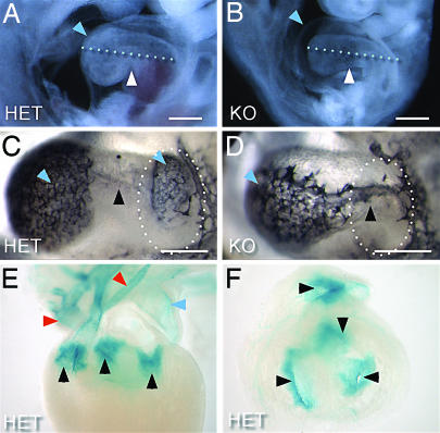
Fig. 5.
Cardiac defects and reporter gene expression in VE-PTPLz/Lz embryos. (A and B) Freshly dissected E9.5 embryos. VE-PTPLz/Lz embryos have pericardial edema (blue arrow) (B). Although knockout embryos are smaller and developmentally delayed compared with their VE-PTPLz/+ littermates (A), their hearts tend to have similar lengths (dotted line is the same length in A and B). (C and D) Whole-mount PECAM-1 staining of E9.5 hearts. The pattern in the VE-PTPLz/+ heart (C) shows distinct ventricle and atrium staining (blue arrows), as well as staining in the connecting atrioventricular canal (black arrow). In the VE-PTPLz/Lz heart (D), there is only staining in the ventricle (blue arrow) and atrioventricular canal (black arrow), but not in the atrium (dotted circle). (E and F) Whole-mount β-gal staining in E15.5 VE-PTPLz/+ heart, showing expression in arteries (red arrows), veins (blue arrows), and high expression in all heart valves (black arrows). Heart valves also shown in thick cross section (F).

