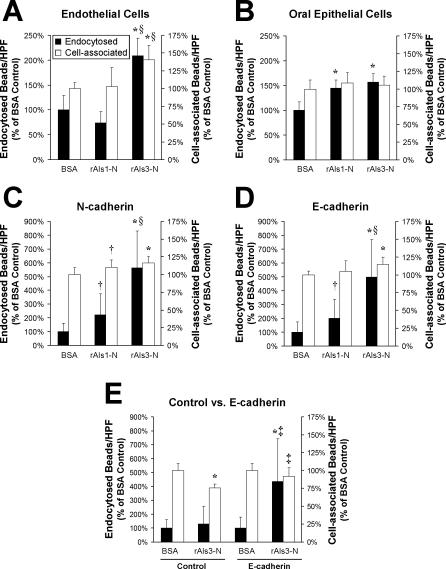
Figure 6. Adherence and Endocytosis of Latex Beads Coated with the N-Terminal Portion of Als1 or Als3.
(A–D) Latex beads were coated with BSA, rAls1-N, or rAls3-N. They were incubated for 45 min with endothelial cells (A), FaDu oral epithelial cells (B), CHO cells expressing human N-cadherin (C), or CHO cells expressing human E-cadherin (D), after which the number of endocytosed and cell-associated beads was determined.
(E) The interactions of BSA- and rAls3-N–coated beads with control CHO cells expressing no human cadherins (Control) were compared with those of CHO cells expressing human E-cadherin (E-cadherin) by the same method as in (A–D). Data are expressed as a percentage of the results with beads coated with BSA and are the mean ± SD of three or four experiments, each performed in triplicate. *, p ≤ 0.005 compared to beads coated with BSA; †p < 0.05 compared to beads coated with BSA; §p < 0.005 compared to beads coated with rAls1-N; and ‡p ≤ 0.01 compared to control CHO cells.

