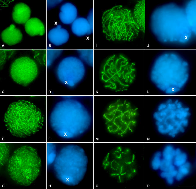
Figure 2. SMC3 Location in Spermatogonial Cells and in First Meiotic Prophase in the Grasshopper L. migratoria Spermatocytes.
(A and B) SMC3 is uniformly scattered throughout the nuclei of the spermatogonial cells, with the exception of the periphery of the single X chromosome. Note that the X chromosome is located into a nuclear protrusion.
(C and D) In this preleptotene spermatocyte, SMC3 appears forming thin cohesion axis threads. Note the absence of labeling in the X chromosome.
(E and F) In leptotene, there is a continuous formation of thin and single SMC3 treads, uniformly distributed into the entire nucleus. Note that the width of the X chromosome SMC3 axis is similar to those present in the autosomes.
(G and H) The onset of zygotene in grasshopper spermatocytes is characterized by the presence of one or two regions of synapsis initiation. These synapsed regions are clearly seen due to the doubleness of the SMC3 threads width.
(I and J) Zygotene spermatocyte in which the “bouquet” configuration is evident.
(K and L) Pachytene spermatocyte characterized by the complete synapsis of autosomes denoted by the double width of the SMC3 lines. A thinner axial signal is present in the unsynapsed X chromosome univalent.
(M and N) Early diplotene, in which desynapsis is accompanied by a barbed wire–like aspect of the SMC3 lines.
(O and P) Late diplotene spermatocyte in which SMC3 is located at the interchromatid domain. It is interesting to note that no signals are found at the centromere regions. (B, D, F, H, J, L, N, and P) correspond to the DAPI-stained chromatin of the spermatocytes. The position of the single sex chromosome is marked with an X. 3-D reconstructions of all the cells of this plate are available in Video S1–S8.

