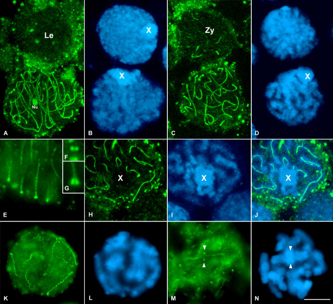
Figure 3. SA1 Location in the Grasshopper E. plorans First Meiotic Prophase Spermatocytes.
(A and B) Leptotene and pachytene spermatocytes. Note that, despite the fact that this image corresponds to the proyection of several focal planes, there is no SA1 signaling in the leptotene spermatocyte. Le, leptotene; Nu, nucleolus.
(C and D) Zygotene and pachytene spermatocytes. Note the presence of short SA1 threads in the zygotene nucleus. Zy, zygotene.
(E) Magnification of the periphery of a pachytene nucleus. Note the accumulation of SA1 at the ends of the linear structures formed by SA1 at their contact with the nuclear envelope.
(F and G) Enlargements of this association in frontal (F) and lateral (G) views, respectively.
(H and I) Projection of all focal planes throughout the univalent sex chromosome from a pachytene spermatocyte. The chromatin of this chromosome is easily distinguished. Note the absence of SA1 signal inside the sex chromosome.
(J) Merged image of the SA1 staining and the chromatin counterstaining. The presence of SA1 signaling in the autosomes is quite evident.
(K and L) Early diplotene cell in which a barbed wire–like staining of SA1 is seen.
(M and N) Late diplotene spermatocyte showing SA1 staining at the interchromatid domain. Note the SA1 accumulations present at the homologous centromere regions of the bivalents (arrowheads). (B, D, I, L, and N) correspond to the DAPI-stained chromatin of the spermatocytes. The position of the single sex chromosome is marked with an X. (A, B, C, D, H, I, and J) are images from confocal microscopy. (E, F, G, K, L, M, and N) are images from fluorescence microscopy.

