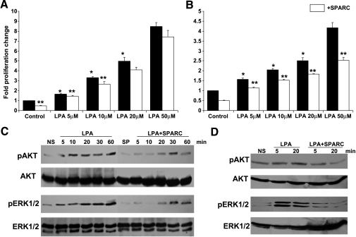
Figure 4.
Effect of SPARC on LPA-induced proliferation and survival signaling in ovarian cancer cells. Cell proliferation of SKOV3 (A) and OVCAR3 (B) cells in response to indicated concentrations of LPA, in the presence (open bars) and in the absence (closed bars) of SPARC, was assessed by measuring the released formazan at A590. Results are expressed as the mean ± SEM of the fold increase in proliferation relative to unstimulated control cells (assigned a value of 1). *P < .05, LPA-stimulated cells compared to control cells and between different concentrations of LPA. **P < .05, SPARC-treated versus matched unstimulated control or LPA-stimulated cells. Represented are the results of one experiment performed in quadruplicate that was representative of two independent experiments. SKOV3 (C) and OVCAR3 (D) cells starved overnight were pretreated with 20 µg/ml SPARC in SFM for 2 hours, followed by stimulation with 50 µM LPA for indicated time points. Western blot analysis of phosphorylated and total ERK1/2 and AKT was performed as described in the Materials and Methods section. Blots represent the results of three independent experiments. NS = not stimulated; SP = SPARC-treated.

