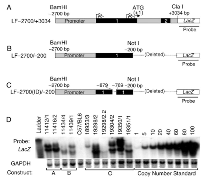
Figure 1. Generation of transgenic mice harboring human Lef-1-promoter fragments driving a LacZ reporter gene.

(A) Diagram of transgene fragment used to generate LF−2700/+3400 transgenic lines. This is the longest fragment of the Lef-1 gene used that contains exons 1 and 2 (shaded black), all of intron 1, and part of intron 2. The closed arrow denotes the position of the start methionine codon (+1) for the Lef-1 protein and open arrows denote the positions of four previously identified transcriptional start sites. The 3′ end of intron 2 contains a splice-acceptor site that is in frame with the LacZ transgene. (B) Diagram of the transgene fragment used to generate LF−2700/−200 transgenic lines. (C) Diagram of the transgene fragment used to generate LF−2700(ID)/−200 transgenic lines. In this construct, a 110 bp fragment encompassing a Wnt response element (−879 to −769) was deleted. (D) Southern blot analysis of all transgenic lines that expressed β-galactosidase at any time during development and which were analyzed for expression patterns at the cellular level in vibrissa and hair follicles. Genomic DNA was digested with BamHI, and Southern blots were sequentially hybridized with a LacZ and GAPDH probes. GAPDH served as an internal control for DNA loading and plasmid copy number controls were spiked into transgene-negative genomic DNA to determine transgene copy number in each line. All founders were determined to have single integration sites based on identical Southern banding patterns in all F1 and F2 generations.
