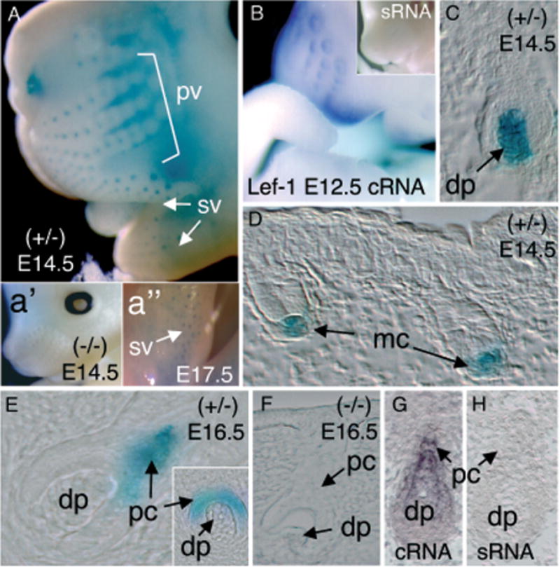
Figure 2. Expression of the LacZ transgene in developing vibrissa follicles of the LF−2700/−200 transgenic lines.

(A) Whole-mount X-gal staining of whiskers in an E14.5 transgenic embryo demonstrating positive LacZ staining in primary vibrissa follicles (pv) and supermumerary vibrissa (sv). Insets depict a lack of X-gal staining in transgene-negative littermates (a′) and persistent supermumerary-vibrissa staining in E17.5 embryos (a″). (B) Whole-mount in situ hybridization for Lef-1 mRNA using an anti-sense (cRNA) probe in an E12.5 embryo. Lef-1 mRNA was detected in vibrissa follicles and lips. No staining was observed using a sense RNA (sRNA) Lef-1 probe (inset of B). (C, D) Histologic analysis of X-gal staining in sections from E14.5 embryos demonstrating predominant Lef-1-LacZ expression in mesenchymal condensates (mc) and dermal papilla (dp) of early stage vibrissa follicles. (E) In contrast to LacZ expression observed at E14.5, Lef-1-LacZ expression was observed in the epithelial-derived pre-cortex (pc) of vibrissa in E16.5 transgenic embryo. (F) No X-gal staining was observed during vibrissa development in transgene-negative littermates at any of the time points evaluated. (G) In situ hybridization using a Lef-1 cRNA probe demonstrates Lef-1-mRNA expression in the differentiating pre-cortex (pc) and dermal papilla (dp) of vibrissa in E14.5 mouse embryos. (H) No hybridization was observed in vibrissa following hybridization of E14.5 embryos with the sense RNA (sRNA) Lef-1 probe.
