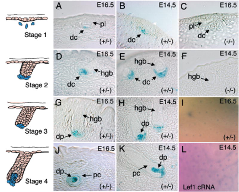
Figure 3. Comparative expression of the Lef-1 promoter during early stage vibrissa and hair-follicle morphogenesis in the LF−2700/−200 transgenic lines.

The left-hand column depicts schematic drawings of early stages of vibrissa/hair-follicle morphology (stages 1–4) (Paus et al, 1999). Photomicrographs of X-gal-stained sections are shown for each corresponding developmental stage. Representative hair follicles (A, C, D, G, and J) and vibrissa follicles (B, F, E, H, and K) from each developmental stage are shown for the given genotypes (C and F are transgene-negative controls). The embryonic stage from which each section was derived is also given in the upper right-hand corner of the figure. (I) Whole-mount X-gal staining of E16.5-transgenic mouse skin and (L) whole-mount in situ hybridization of skin using a mouse Lef-1-cRNA anti-sense probe. dc, dermal condensates; dp, dermal papilla; pc, pre-cortex; and hgb, hair germ bud; pl, placode.
