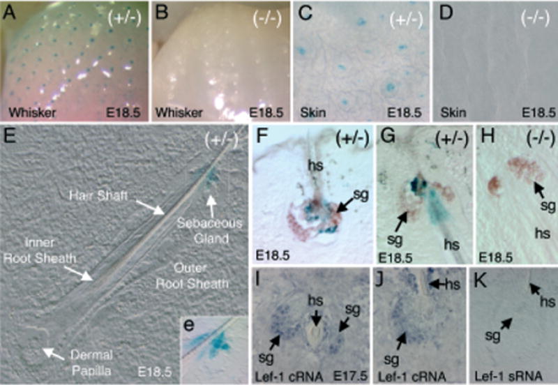
Figure 4. Expression of the LF−2700/+ 3034 Lef-1 promoter in sebaceous glands.

(A–D) Whole-mount X-gal staining of the whisker pad (A, B) and skin (C, D) of transgenic (A, C) and non-transgenic (B, D) embryos at E18.5. (E) Nomarski photomicrograph of a whisker cross-cryosection from a transgene-positive animal; the various structural features are annotated. The X-gal staining in the sebaceous gland is more obviously displayed in the bright field inset (e) of the enlarged boxed regions (F–H). Oil Red O-stained sections of E18.5 embryo whiskers also stained in X-gal. Sebaceous glands in whiskers stained brown using Oil Red O (arrows). (F) A cross-section and (G) sagittal-section of whiskers demonstrates a cluster of X-gal-positive cells surrounding the hair shaft in the center of the sebaceous gland from transgene-positive embryos. (H) No X-gal-positive staining was observed in whisker sebaceous glands of transgene-negative embryos. (I–K) In situ hybridization of E17.5 whiskers using anti-sense (I, J) or sense (K) mouse Lef-1-RNA probes. Lef-1 mRNA is expressed in a subset of cells within sebaceous glands (sg) surrounding the hair shaft (hs).
