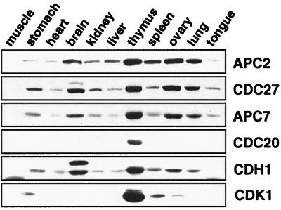
Figure 1.
Analysis of APC expression in mouse tissues. Equal amounts of protein from 10,000 × g supernatant fractions from different mouse tissue extracts were separated by SDS/PAGE and analyzed by immunoblotting by using antibodies to the indicated proteins. The CDH1 crossreactive band of slower mobility detected in brain extract neither cofractionated nor coimmunoprecipitated with APC (see Fig. 4B and not shown).

