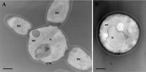
FIG. 4.
Transmission electron micrographs of C. glabrata isolates 21229 (A) and 21231 (B). Transmission electron microscopy confirmed the pseudohyphal growth of isolate 21229, with cells presenting up to three daughter cells of similar size, and revealed the ultrastructural changes of their cell wall with a thinner inner layer compared with cells of control isolate. N, nucleus; mt, mitochondrion; CW, cell wall; DC, daughter cell.

