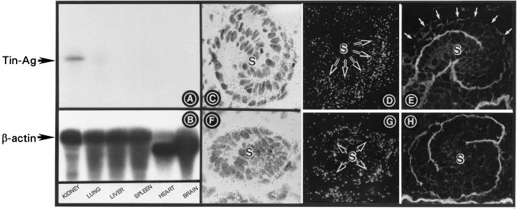
Figure 2.
(A and B) Northern blot analyses of TIN-ag and β-actin. A ≈2.0-kilobase transcript is observed in the kidney, and a very faint band is observed in the lung. The β-actin mRNA expression is similar in all the tissues. (C and D) TIN-ag mRNA expression in the S-shaped body (S). (E) TIN-ag protein expression. The expression in the basal lamina is confined to the distal convolution and is absent in the upper convolution (white arrows). (F and G) HS-PG mRNA expression. (H) HS-PG protein expression is confined to the basal lamina of both the convolutions. (C–H, ×250.)

