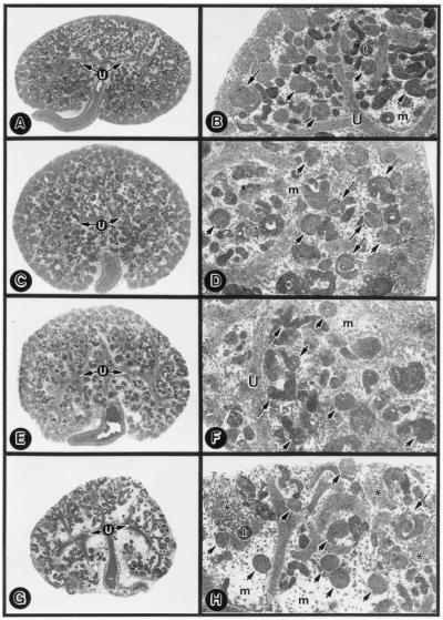
Figure 6.
Low- (A, C, E, and G) and high-magnification (B, D, F, and H) micrographs of the goat-IgG-treated (A and B, 10 μg/ml) and anti-TIN-ag antibody-treated (C and D, 2.5 μg/ml; E and F, 5.0 μg/ml; G and H, 7.5 μg/ml) E13 explants. Normal goat-IgG induced no alterations in metanephroi. Anti-TIN-ag antibody treatment induced a dose-dependent deformation of the S-shaped bodies (long arrows) and reduction of the tubules (t), whereas glomeruli (short arrows) were unaffected. A few patches of compacted mesenchyme (asterisks) are discernible. u, ureteric bud branches; m, metanephric mesenchyme. (A, C, E and G, ×20; B, D, F, and H, ×80.)

