
Fig. 2.
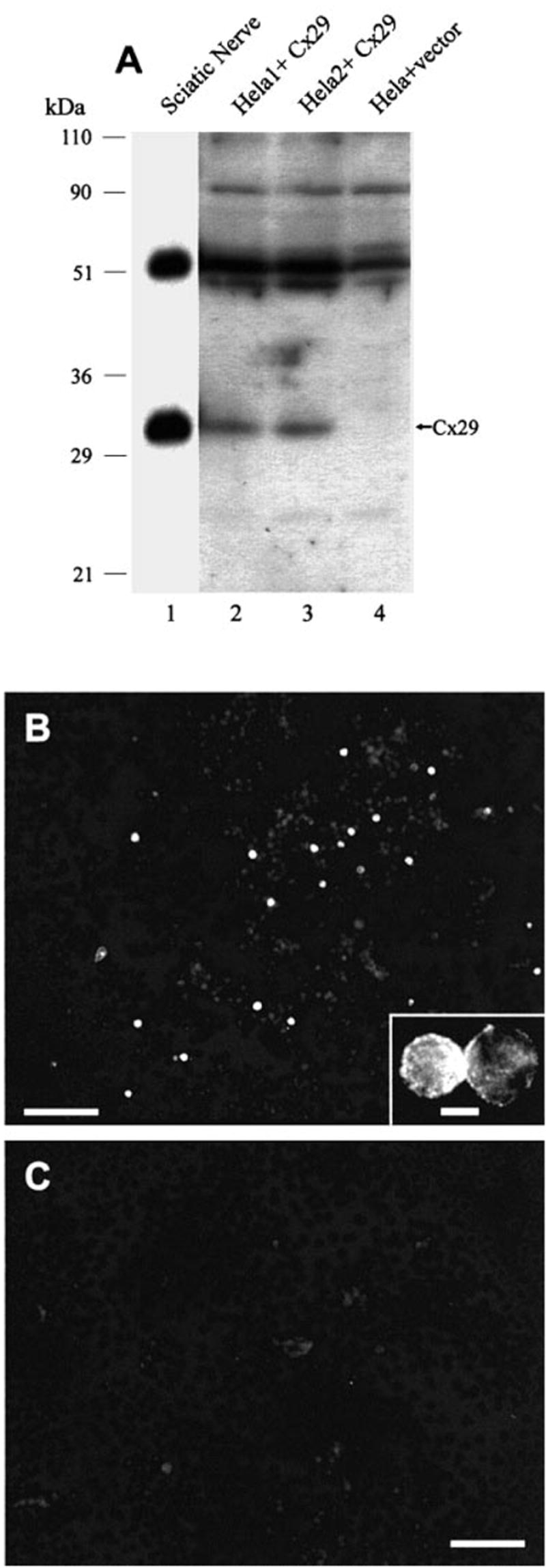
(A) Western blots of Cx29 in HeLa cells transiently transfected with Cx29 cDNA. Lysates from two separate cultures of cells transfected with Cx29 expression vector show antibody detection of monomeric Cx29 (lanes 2 and 3) corresponding to that of Cx29 in homogenate of sciatic nerve (lane 1), while no detection is seen in control HeLa cells transfected with empty vector (lane 4). (B and C) Immunofluorescence of Cx29 in transfected HeLa cells showing robust immunolabelling in a small percentage of cells (B), and total absence of labelling in cells transfected with empty vector (C). Inset shows immunofluorescence within a pair of cells and around their periphery. Scale bar, 100 μm (B and C), 25 μm (inset).