
Fig. 3.
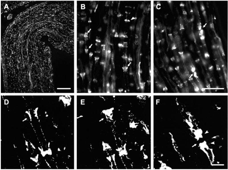
Immunofluorescence of Cx29 in sections of sciatic nerve from CD1 mice. (A) Low magnification showing distribution of labelling with anti-Cx29 antibody. (B and C) Magnifications showing labelling of either conical structures (B, arrows) or bands (B, arrowhead) orientated perpendicular to the long axis of fibers, and accumulation of staining at nodes of Ranvier (C, arrows). (D and E) Laser scanning confocal micrographs of the same field at different planes of focus showing collar of immunolabelling midway through a fibre (D, arrow) and closer to its surface (E, arrow). Note absence of labelling within the axon in D. (F) Confocal micrograph showing accumulation of labelling for Cx29 on each side (arrowheads) of a node of Ranvier (arrow). Scale bars, 100 μm (A); 25 μm (B and C); 5 μm (D–F).