
Fig. 9.
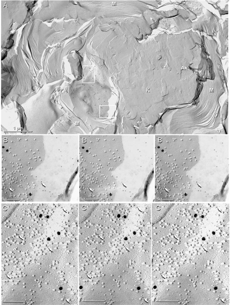
IMP rosettes and clusters in innermost myelin layer in sciatic nerve of Cx32 KO mouse after double-labelling for Cx29 (6 and 18-nm gold) and Cx32 (12-nm gold; none present). Label for Cx29 is abundant beneath P-face IMPs in the innermost layer of myelin (A, inscribed boxes), but was absent in other layers of myelin (A, lower left) or in areas of knife scrape (K) (A, centre-right). (B) Magnification of lower box in A. The 6-nm gold beads (arrows) are more easily seen in areas where shadowed debris prevented platinum deposition. In the area not coated with platinum, IMPs are faintly delineated by the very thin, rotary-deposited carbon film. (C) Magnified upper box in A showing clustered IMPs labelled with six 18-nm and 24 6-nm gold beads (arrows). The 6-nm gold beads are more readily evident by first viewing the intaglio images (right two images), then the true stereoscopic perspective (left two images). Scale bars, 1 μm (A); 0.1 μm (B and C).