Figure 2.
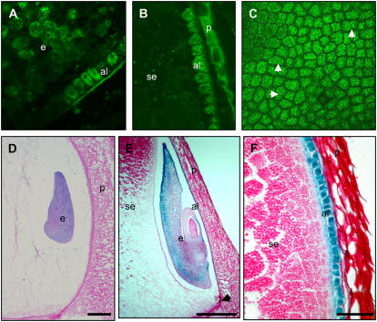
Regulation of reporter genes by the Vp1 promoter in transgenic maize. A to C, Vp1∷GFP expression in 10-DAP kernels analyzed by confocal laser-scanning microscopy. A, Embryo (e) and aleurone (al) cells expressing GFP (denoted by bright-green, grainy appearance). B, Vp1∷GFP expression in aleurone cells only (note autofluorescence of the pericarp tissue). C, GFP expression in an aleurone peel. White arrowheads indicate newly dividing cells. D to F, Vp1∷GUS expression (denoted by purple/blue precipitate) in developing maize seeds at 7 (D) and 10 (E and F) DAP. GUS expression is detected in the transition embryo from around 7 DAP (shown in D). E, GUS staining is detectable at higher levels in the scutellum at later stages of embryogenesis (indicated by arrows), as well as in the endosperm aleurone (shown here on the germinal kernel face and extending down until the basal endosperm transfer region, indicated by arrowhead). F, Cells of aleurone and subaleurone (sa) layers staining for GUS. Ten-micrometer-thick sections were stained for GUS, counterstained with periodic acid Schiff reagent (crimson color), and visualized under bright-field microscopy. Scale bars = 25 μm (D and E); 10 μm (F).

