Figure 3.
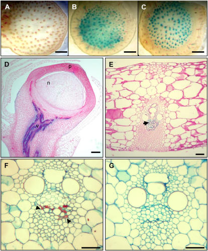
Stress-induced Vp1∷GUS transgene and endogenous gene expression in maize vegetative tissues. A to C, GUS-stained hand-cut transverse sections of ear shank tissue from plants carrying the Vp1∷GUS reporter gene. Water control (A), 6-h desiccation (B), and 6-h incubation in 0.6 m Suc (C). GUS expression is only detected in the phloem. Sections were counterstained with phloroglucinol-HCl for detection of lignin (denoted by red precipitate). D to E, Vp1∷GUS expression after drought stress treatment in the vascular tissue (v) of an ear pedicel (D) and transverse leaf section (E). GUS staining is localized in the phloem parenchyma cells (ph), indicated by arrow. F and G, Vp1 mRNA in situ localization after drought stress treatment in phloem companion cells (denoted by red/purple staining and indicated by black arrowheads) in transverse sections of stems imaged under bright-field optics after saffranin fast-green staining. F, Antisense probe. G, Sense probe. Scale bars = 50 μm (A–C); 25 μm (D); 5 μm (E–G).

