
Figure 13.
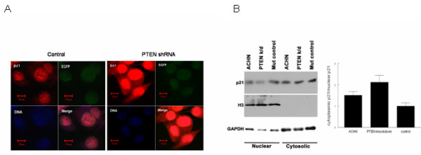
PTEN attenuation enhances p21 expression and cytosolic localization. a. ACHN cells stably transfected with PTEN-shRNA or empty vector were immunostained with anti-p21 antibody combined with Alexa Fluor® 594-conjugated secondary antibody for detection under UV microscopy. DAPI was used to demarcate nuclei, and EGFP (expressed by the plasmid) was used as a transfection control. b. Cells were fractionated for immunoblotting, with antibodies against mono methyl Histone H3-Lys4 and GAPDH used as nuclear and cytosolic controls, and p21 levels were quantified using densitometry. The ratio of cytosolic/nuclear p21 is shown graphically; the error bars indicate intra-experimental variation (standard deviation) from two replicates.