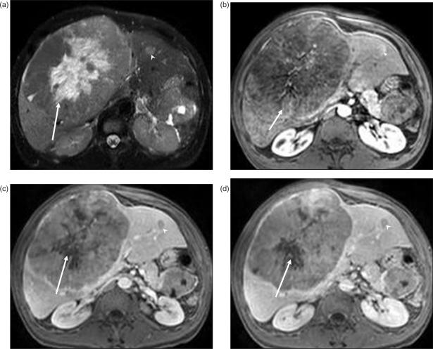
Figure 2.
Islet cell tumor metastases. (a) T2W image demonstrates a well-circumscribed large mass replacing the right lobe of the liver. The central (arrow) hyperintense area represents necrosis as the metastases has rapidly outgrown its blood supply. Post-contrast T1W images reveal heterogenous enhancement of the peripheral viable portion of the metastases in arterial phase (arrow in (b)). In portal venous (c) and delayed phase (d), the lesion is iso- to hypo-intense to the liver, with the central necrotic portion (arrow) remaining unenhanced. Another small metastatic lesion (arrowhead) is seen in the left lobe of the liver.

