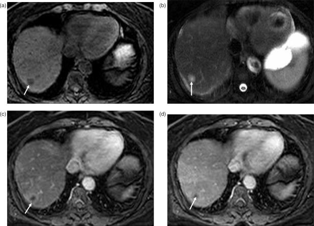
Figure 5.
Hypovascular liver metastases from pancreatic adenocarcinoma. (a) T1W image shows hypo-intense lesion (arrow) in segment VII of the liver, which is hyper-intense (arrow) on the T2W image (b). (c) Lesion demonstrates subtle concentric perilesional enhancement (arrow) on post-contrast arterial phase image. (d) On portal venous phase image perilesional enhancement becomes iso-intense to background liver and the center of the lesion (arrow) does not enhance due to necrosis.

