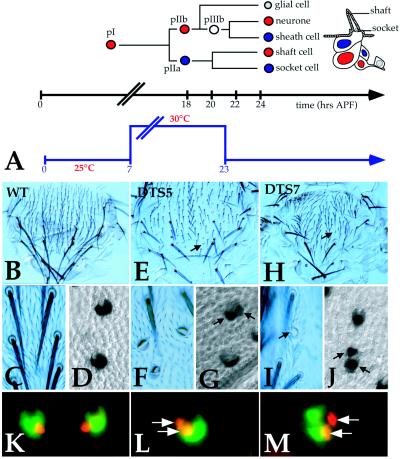
Figure 1.
Interfering with proteasome function in early pupae yields a double-socket bristle phenotype. (A) Diagram of the bristle lineage in the notum. pI divides asymmetrically to generate pIIa and pIIb. Notch signaling is active in pIIa (blue cell) and inhibited by Numb in pIIb (red cell). pIIb divides to generate pIIIb (white cell) and a small glial cell (gray cell) that will migrate away soon after pIIIb division. The divisions of the pIIa and pIIIb cells give rise to the four mechanosensory organ cells, schematically shown on a diagram of a differentiated microchaete. Notch signaling is active in the sheath and socket cells (in blue) and blocked by Numb in the neuron and shaft cells (in red). The approximate timing of these divisions is indicated by the black time scale below. APF, after puparium formation. The temperature shifts applied to DTS5 and DTS7 pupae are shown below as a blue time scale bar. (B–J) Cuticular preparations of dorsal thoraces from pharate adults (panels B, C, E, F, H, and I) and nota dissected from 24-hr-APF pupae stained for Su(H) immunoreactivity (horseradish peroxidase staining in D, G, and J). Wild-type (B–D), DTS5/+ (E–G), and DTS7/+ (H–J) were heat-pulsed at 30°C during a 7- 23-hr-APF time period as indicated in A. No bristle phenotype was seen at the permissive temperature (not shown). Arrows point to double-socket bristles (E, H, and I) and pairs of socket cells (G and J). (K–M) Indirect immunofluorescence staining of nota dissected from 28-hr-APF wild-type (K), DTS5/+ (L), and DTS7/+ (M) pupae that were heat-pulsed at 29°C at 0–28 hr APF. Duplicated sheath cells in DTS5 and DTS7 mutants are shown by white arrows. Each panel correspond to the merge of images from two focal planes: the socket cells (in green: Suppressor of Hairless) were found in the epithelial plane, whereas the sheath cells (in red: Prospero) were subepithelial. These Prospero-positive cells cannot correspond to glial cells for several reasons: first, at this late stage, no glial cells accumulate Prospero; furthermore glial cells migrate away from microchaetes around 23–24 hr APF and are no longer associated with microchaetes at 28 hr APF; finally, glial cell nuclei are distinctly smaller than sheath cell nuclei (7). In all panels anterior is up.

