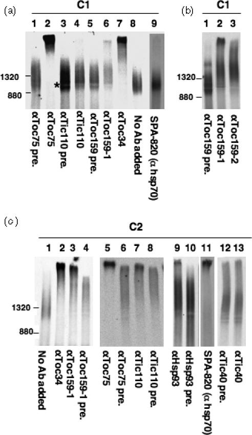
Figure 4. Antibody-shift BN-PAGE analyses of the composition of the C1 and C2 complexes.

(a) The sucrose-density gradient fraction containing C1 was incubated with various antibodies, as labeled at the bottom, to remove possible components in C1 (pre., pre-immune serum of the respective antibody). The sharp band between 880 and 1320 kDa was formed due to the presence of a major serum protein at the position marked by the asterisk.
(b) Same as in (a) and the experiment was repeated with a second anti-Toc159 serum (αToc159-2).
(c) The sucrose-density gradient fraction containing C2 was incubated with various antibodies, as labeled at the bottom, to remove possible components in C2.
