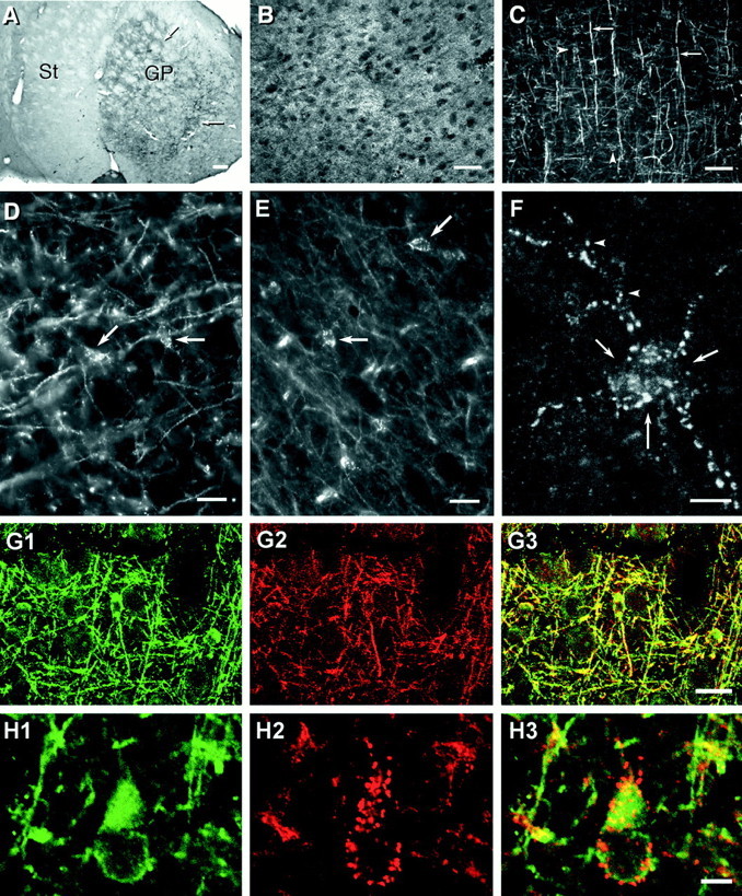
Fig. 2.

Photomicrographs showing patterns of immunolabeling obtained with various anti-connexin antibodies used in double-immunofluorescence and FRIL studies. A, Low magnification of immunoperoxidase labeling (arrows) with a monoclonal anti-Cx30 antibody. The section shows dense punctate labeling in the globus pallidus (GP) and weaker labeling in the striatum (St). B, Cerebral cortex showing a high concentration of punctate immunofluorescence with anti-Cx43 antibody 18A. Dark ovals represent unstained neuronal cell bodies. C, Cx32 labeling of myelinated fibers (arrows) as well as oligodendrocyte cell bodies (arrowheads; cells barely discernable at this magnification) using antibody 7C7. D, E, Higher magnifications showing Cx32-positive puncta with antibody 7C7 along the surface of oligodendrocytes in the cerebral cortex (D; arrows) and ventral lateral nucleus of the thalamus (E; arrows). F, Through-focus confocal micrograph of Cx32-immunopositive puncta distributed on an oligodendrocyte soma (arrows) and its processes (arrowheads). G, Double immunofluorescence of the oligodendrocyte marker CNPase (G1, green) and Cx32 (G2,red) in the same field of cerebral cortex. CNPase-positive cells are also immunopositive for Cx32 as seen by theoverlay of images (G3,yellow). H, Higher magnification double-immunofluorescence confocal micrographs showing a CNPase-positive oligodendrocyte in cerebral cortex (H1) decorated with numerous Cx32-immunopositive puncta (red puncta in H2 and red andyellow puncta in the overlay of images inH3). Scale bars: A, 200 μm; B, C, 50 μm; D, E, G, 20 μm; F, H, 5 μm.
