
Figure 3.
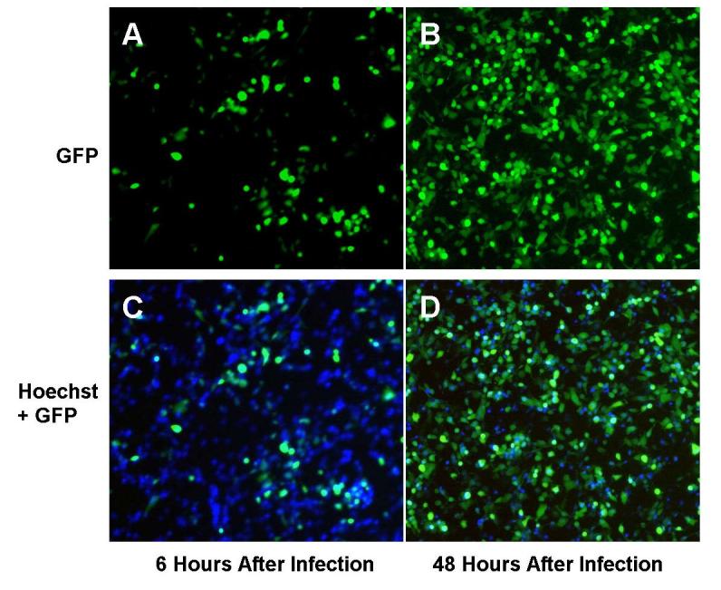
In vitro vector spread as identified by GFP expression. Following treatment with NV1066, infected MSTO-211H cells express GFP, as determined by fluorescent microscopy. GFP expressing cells increased over time, to nearly 100% at all MOIs, indicating that all the cells were infected by NV1066. Malignant mesothelioma cells were infected by NV1066 and their nucleus was stained with Hoechst staining (blue). Images were taken under DAPI filter (to identify individual cells by blue nuclear staining) and GFP filter (to identify GFP expressing infected cells) at 6 and 48 hours. Images were overlapped using metamorph software. Over time, NV1066 was able to infect, replicate, propagate, and express GFP in all malignant mesothelioma cells. MOI = Multiplicity of infection, ratio of viral particles to tumor cells, GFP = Green fluorescent protein.