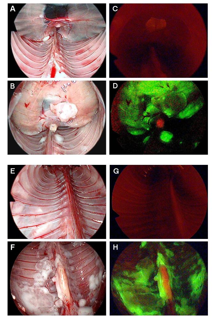
Figure 5.
Thoracoscopic identification of diaphragmatic and pleural mesothelioma. In vivo, viral uptake and GFP expression in pleural cavities and diaphragmatic mesothelioma can be easily identified by using a thoracoscope with GFP filter. All the mice were injected with NV1066 intrapleurally. GFP expression can be visualized selectively only in malignant mesothelioma tissue as early as 48 - 72 hours. Normal mouse diaphragm (A), diaphragmatic mesothelioma (B) were examined under bright-field. Examination under GFP mode identifies no GFP expression in normal diaphragm (C) and selective infection of diaphragmatic mesothelioma by NV1066 (D). Normal parietal pleura (E), pleural malignant mesothelioma (F) was identified under bright-field. No GFP expression in normal parietal pleura (G) and selective infection of pleural mesothelioma (H) was identified by thoracoscope fitted with fluorescent filters for GFP. GFP = Green fluorescent protein.

