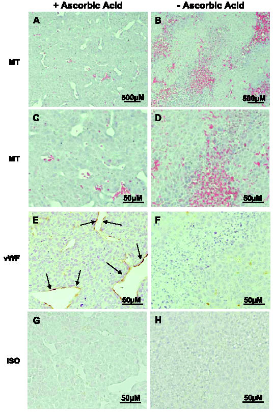
Figure 3.

Tumors in ascorbic acid-restricted Gulo-/- mice exhibit poorly formed vasculature with multiple hemorrhagic foci. Tissue sections from representative tumors in replete (A, C, E, and G) and depleted (B, D, F, and H) Gulo-/- mice were examined for vasculature after staining with Masson's trichrome (MT) stain (A and C), anti-vWF polyclonal antibody (vWF; E and F), or preimmune rabbit serum (ISO; G and H). Endothelial cells stained with α-vWF are in brown and shown by arrows (A and B: original magnification, x200; C–H: original magnification, x400).
