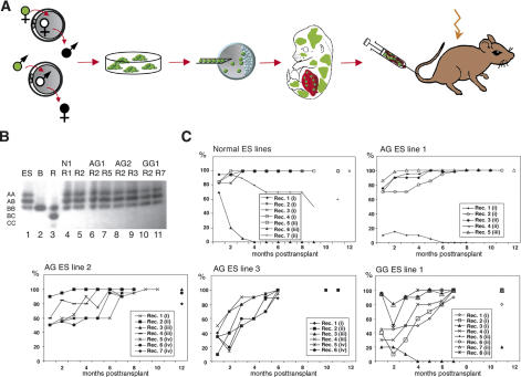
Figure 1.
Multilineage reconstitution by androgenetic and gynogenetic cells. (A) Experimental design. EGFP-expressing ES cell lines derived from androgenetic and gynogenetic embryos produced by pronuclear transfer between zygotes were injected into blastocysts. After embryo transfer, fetuses were recovered at 13.5–14.5 dpc, chimeras were identified by eGFP-fluorescence, and fetal liver from chimeras was transplanted into lethally irradiated recipient mice. (B) Analysis of GPI-1 isoenzymes to identify contribution of ES cell-derived cells to the peripheral blood of recipients. Lanes 1–3 show the GPI-1 isoenzyme dimers present in the ES cells ([ES] A and B isoforms), blastocysts ([B] B isoform only), and adult recipients ([R] B and C isoforms), respectively. (GPI-1 forms homo- and heterodimers, such that cells containing A and B isoforms contain AA, AB, and BB dimers; all dimers are indicated on the left.) Lanes 4–11 show the predominance of ES cell-derived cells (A, B isoforms) in the peripheral blood of individual recipients (R) 6–8 mo after transplantation of ES cell chimeric fetal liver (ES line indicated on top). (C) Presence of ES cell-derived cells in peripheral blood of recipients over time as determined by GPI-1 analysis. Numbers in parentheses indicate transplanted fetal liver cell pools, with identical numbers referring to the same pool.

