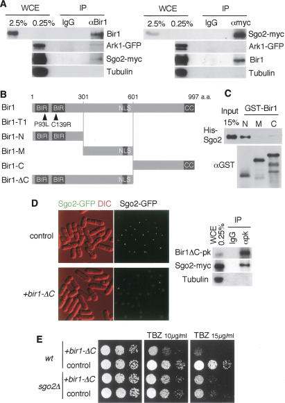
Figure 6.
Sgo2 associates with Bir1. (A) Coimmunoprecipitation of Bir1 and Sgo2. Cells expressing Sgo2-myc and Ark1-GFP were arrested at prometaphase by incubating for 10 h at 20°C (by nda3-KM311) and cross-linked by 0.8% formaldehyde. Immunoprecipitates (IP) from cell extracts obtained by anti-Bir1 or anti-myc antibody as well as control IgG were analyzed by Western blot using anti-Bir1, anti-GFP, anti-myc, and anti-tubulin (control) antibodies. (WCE) Whole-cell extract used for immunoprecipitation. (B) Schematic drawing of the indicated mutant or truncated constructs of Bir1. (C) GST-Bir1-N, GST-Bir1-M, GST-Bir1-C, and His-Sgo2 recombinant proteins were expressed and purified from E. coli. His-Sgo2 was incubated with the indicated GST-tagged proteins bound to beads. Bead-bound fractions were separated by SDS-PAGE and analyzed by Western blotting using antibodies against His and GST. (D) sgo2+-GFP cells carrying a vector or plasmid expressing Bir1-ΔC were examined for the localization of Sgo2-GFP. Cell extracts prepared from sgo2+-myc cells expressing Bir1-ΔC tagged with pk were immunoprecipitated using control IgG and anti-pk antibody and detected by Western blot using anti-pk, anti-myc, and anti-tubulin (control) antibodies. (E) Wild-type or sgo2Δ cells expressing Bir1-ΔC or vector alone were spotted onto minimal medium plates containing 0, 10, or 15 μg/mL TBZ and incubated at 30°C.

