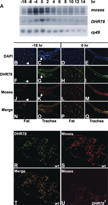
Figure 4.
Spatial and temporal expression profiles of Moses and DHR78. (A) Northern blot analysis to detect moses and DHR78 transcripts of w1118 animals staged as mid-L3 (−18 and −8 h), late L3 (−4 h), newly formed prepupae (0 h), prepupae staged at 2-h intervals (2, 4, 6, 8, and 10 h), and pupae (12 and 14 h). Hours are relative to puparium formation. Hybridization to detect rp49 mRNA was included as a control for loading and transfer. (B–Q) Fat body and trachea from either −18-h mid-L3 or 0-h prepupae were stained with DAPI (B–E), DHR78 antibodies (F–I), or Moses antibodies (J–M). (N–Q) A merge of the antibody patterns. White arrows mark nuclei that contain DHR78 and Moses protein. (R–T) Antibody stains of giant salivary gland polytene chromosomes from late L3 (−4 h) w1118 animals (wt) to detect DHR78 (R), Moses (S), and the merge of the two (T) bound to chromatin. (U) Antibody stains of Moses in hs-DHR78/hs-DHR78; DHR782/DHR782 mutants that have been rescued to late L3 (−4 h) by transient expression of a DHR78 transgene during embryogenesis for 30 min at 37°C and maintained at 18°C through development, showing loss of chromatin localization.

