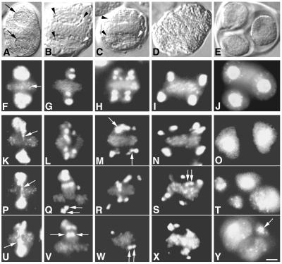
Figure 5.
Wild-type and mutant meiosis II. Images of similar stages are shown in the same column. (A–E) DIC images of wild-type MMCs. (F–J) DAPI images of the cells shown in A–E, respectively. (K–Y) DAPI fluorescent images of ask1-1 MMC s. DIC images of ask1-1 cells (not shown) are abnormal and not well defined. Therefore, assignment of stages for ask1-1 cell relies largely on DAPI staining patterns. (A) Prophase II, with two nuclei (arrows). (B) Metaphase II, with two spindles; the arrowheads mark the positions of the spindle poles. (C) Anaphase II, with two spindles are marked as in B. (D) Telophase II, with spindles no longer present. (E) A tetrad, showing three of the four spores; the nuclei are visible. (F) Prophase II, with an organelle band (arrow) and two brightly stained groups of chromosomes. (G) Metaphase II, with two groups of highly condensed chromosomes, one on each side of the organelle band; only some chromosomes are visible in this plain of focus. (H) Anaphase II. The chromatids had separated, moving apart along the axis of the spindle. (I) Telophase II. Four brightly stained areas represent four groups of partially decondensed chromosomes. (J) Three stained nuclei of the tetrad. (K, P, and U) Three ask1-1 cells at prophase II. Two brightly DAPI-staining regions were seen on either side of the organelle band, and some DAPI-staining materials were found across the organelle band (arrows). (L) A cell at metaphase II; the chromosomes were not localized to two regions. Some were near the organelle band. (Q and V) Two images of the same metaphase II cell at different focal planes. The area above the organelle band had much more staining than that below the band. (M) Approximately anaphase II. DNA seemed to be extended (arrows) parallel to the organelle band. (R and W) Two images of the same cell at approximately anaphase II, showing scattering of chromosomes. (N) Approximately telophase II, showing six regions of condensed chromosomes. (S and X) Two images of the same cell showing several regions of condensed chromosomes. Some chromosomes overlapped the organelle band. (O) A tetrad with two spores. (T) A tetrad with four spores, two of them are larger than the other two. (Y) An image of a tetrad showing three of its five spores. One of them (arrow) is extremely small. Double arrows in Q, S, V, and W indicate adjacent chromosomes. All panels have the same magnification. (Scale bar = 5 μm.)

