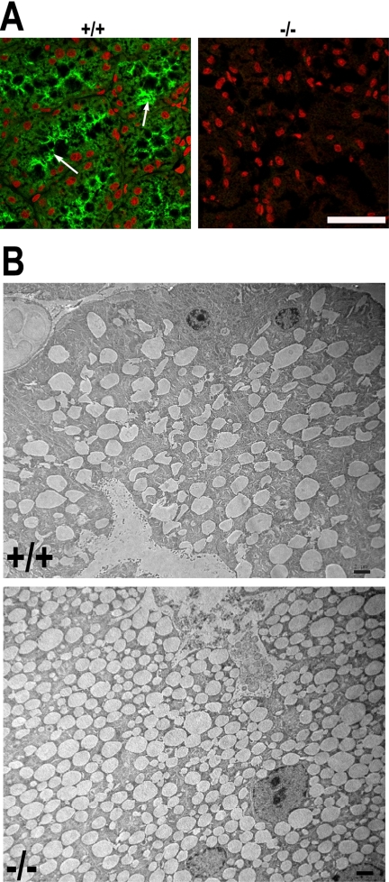
Figure 4.
Ablation of VAMP8 led to accumulation of secretory granules in lacrimal gland acinar cells. (A) Immunofluorescence staining showing VAMP8 expression in the apical region of lacrimal gland acinar cells. VAMP8 was stained green, whereas the nuclei are red. Arrows indicate apical poles of acinar cells. Scale bar, 50 μm. (B) Electron microscopy showing accumulation of secretory granules in VAMP8-null acinar cells of lacrimal glands. Scale bar, 2 μm. +/+, wild-type; −/−, VAMP8-null.

