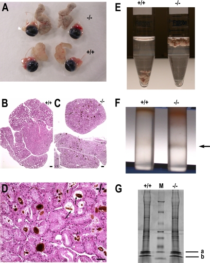
Figure 5.
Protein deposits (aggregates) in VAMP8-null lacrimal glands. (A) Gross appearance of VAMP8-null (top) and wild-type (bottom) lacrimal glands together with eyeballs. (B) H&E staining of normal lacrimal gland section. (C) H&E staining of VAMP8-null lacrimal gland section showing accumulation of dark-brown deposits. (D) High magnification of an H&E-stained section of VAMP8-null lacrimal glands showing protein deposits in the lumen of ducts and acini. Arrows indicate lumen of the ducts. Scale bar, 50 μm. (E) Photography of lacrimal glands in PBS showing gravity density difference between normal (+/+) and mutant (−/−) lacrimal glands. (F) Sucrose density fractionation of lacrimal gland homogenate showing a dark-gray band at the interface between 2 and 1.5 M sucrose (indicated by an arrow). (G) SDS-PAGE showing protein components in the deposits. From bottom to top, the molecular weights of the protein markers (M) were 6.5, 16.5, 25, 32.5, 47.5, 63, 83, and 175 kDa, respectively. Bands were identified by mass spectrometry as histone 1 and H2b (a) and hemoglobin beta-1 subunit (b).

