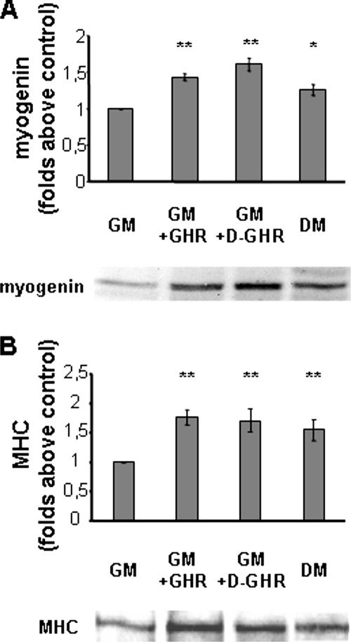
Figure 2.
Western blot analysis of myogenic markers expression. C2C12 myoblasts were incubated in GM for 24 or 72 h with 10 nM GHR or D-GHR. The content of myogenin and MHC was measured by Western blot of whole cell lysates and the intensity of the bands quantified. Cells differentiated in DM were considered as positive control. (A) Myogenin expression. (B) MHC expression. Representative Western blots are shown at the bottom of each panel. **p < 0.01 and *p < 0.05 versus control.

