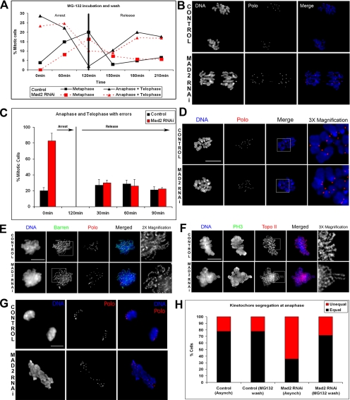
Figure 4.
Reversion of the phenotypes caused by depletion of Mad2. To determine whether the Mad2-associated phenotypes could be reverted by providing additional time in prometaphase/metaphase, S2 cells previously treated with dsRNA against Mad2 for 72 h were incubated for 120 min with a low dose of MG132 (2 μM) and then released from the block by performing a threefold dilution on the cell culture media with fresh media. Samples were then collected every 30 min for immunofluorescence analysis. (A) Quantitative analysis of mitotic progression in control and Mad2-depleted cells before and after reversion. From 0 to 120 min both control and Mad2-depleted cells show a strong decrease in anaphases and telophases and a marked increase in the number of metaphases. After washing MG132 (120 min), the number of metaphases begins to decrease, and the number of anaphase and telophase figures increases. (B) Immunofluorescence shows that 180 min after washing the drug, most anaphases in Mad2-depleted cells do not show chromatin bridges. (C) Quantitative analysis of the anaphase and telophase figures before and after the MG132 treatment shows a complete reversal of the phenotype. (D) Normal chromosomes can be obtained in Mad2-depleted cells if cells are incubated with MG132 to prevent exit from mitosis. Before fixation cells were also treated with colchicine to depolymerize microtubules and induce a better chromosome spread. DNA is shown in blue and the kinetochore marker Polo in red. (E and F) Immunolocalization of essential components for the organization and structure of mitotic chromosomes. Barren and Topoisomerase II in control and Mad2-depleted prometaphases are properly localized to a well-defined sister chromatid axis, in asynchronous cell culture. (G) Analysis of kinetochore segregation in control and Mad2-depleted cells after 72 h of RNAi incubation was carried out on anaphase cells after immunostaining for DNA with anti-Polo antibody. (H) Quantification of chromosome segregation at anaphase shows that in control cells almost 80% of cells segregate kinetochores equally. After depletion of Mad2 nearly 70% of cells also show unequal kinetochore segregation. Providing extra time in prometaphase/metaphase to control cells by incubation in MG132 and then washing out the drug does not alter the frequency of unequal kinetochore segregation. However, a similar treatment in Mad2-depleted cells reduces to almost control levels the frequency of unequal kinetochore segregation. Bar, (D–G) 5 μm. A total of 40 cells were analyzed.

