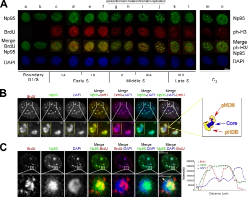
Figure 1.
During mid-S, Np95 targets pericentric heterochromatin and is part of pHDB. (A) Distribution of Np95 during S-phase. NIH-3T3 cells were synchronized in G0. At different time after release, cells were pulse-labeled with BrdU and then stained with anti-Np95 together with either anti-BrdU or anti-pH3 (specific for histone H3 phosphorylated at serine 10). Nuclear counterstaining was visualized with DAPI. (B) Np95 is part of pHDB. NIH-3T3 cells in mid-S phase were pulse-labeled with BrdU and then stained with anti-Np95 together with anti-BrdU. Nuclear counterstaining was visualized with DAPI. Representative pictures are shown. The insets correspond to magnifications of the areas indicated. On the right is a schematic representation of the pHDB of the inset. (C) NIH-3T3 cells in mid-S phase were stained as above and then analyzed by confocal microscopy. Representative pictures are shown. The insets correspond to magnifications of the areas indicated. The histogram shows the local intensity distribution (diagonal white lines through the images) of Np95 in green, BrdU in red, and DAPI in blue.

