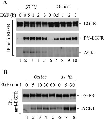
Figure 4.
Determination of the location for the interaction of ACK1 with EGFR. (A) Interaction of endogenous ACK1 with EGFR does not occur on the plasma membrane. The COS7 cells were starved in serum-free medium for 12 h and then placed on ice for 30 min. EGF (100 ng/ml) was added to the cells on ice for the indicated time. The control cells were preincubated and stimulated with EGF (100 ng/ml) at 37°C (lanes 1–5). EGFR was immunoprecipitated with anti-EGFR Mab528 from the cell lysates and immunoblotted with anti-EGFR (1005; top panel). Coimmunoprecipitated ACK1 was detected by immunoblotting with anti-ACKPCC (bottom panel). Tyrosine phosphorylation of EGFR was detected by immunoblotting with anti-PY (4G10; middle panel). (B) Interaction of exogenous ACK1 with EGFR may occur on the plasma membrane. The Myc-tagged ACK1 was transfected into COS7 cells for 36 h followed by serum starvation for 12 h. The experimental procedures, including ice incubation, EGF stimulation, immunoprecipitation, and immunoblotting, were the same as described in A. Top panel, immunoprecipitated EGFR detected by immunoblotting with anti-EGFR (1005); bottom panel, coimmunoprecipitated ACK1 detected by immunoblotting with anti-ACKPCC.

