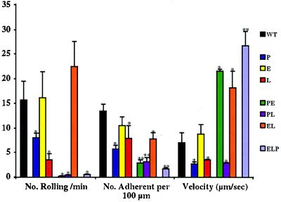
Figure 4.
Leukocyte rolling in TNF-α-stimulated venules. Mice were prepared for intravital microscopy of mesenteric venules 3.5 hr after administration of TNF-α. Venular blood flow and diffractive, rolling leukocytes were videotaped for 20 min. Averages were obtained from at least five animals of each genotype. ∗, P < 0.05; ∗∗, P < 0.01, in comparison to WT mice.

