Fig. 4.
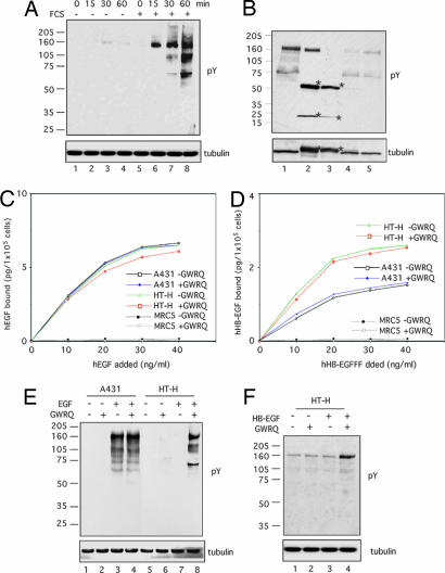
Effect of GWRQ peptide on EGFR activation in HT-H cells. (A) Western blot of tyrosine-phosphorylated proteins. HT-H cells were cultured in medium with or without FCS and were treated with GWRQ peptide at 4°C for 15 min, followed by phosphorylation stimulation at 37°C for a period of 0–60 min. (B) Western blot of tyrosine-phosphorylated proteins immunoprecipitated by a mixture of antibodies against ErbB family EGF receptors. HT-H cells were treated with GWRQ peptide at 4°C for 15 min, followed by an incubation at 37°C for 30 min in the presence of FCS. Samples loaded are as follows: lane 1, total cell lysate; lane 2, immunoprecipitates by a mixture of antibodies for ErbB1, -2, -3, and -4; lane 3, immunoprecipitates by control antibodies; lane 4, supernatant of immunoprecipitates by a mixture of antibodies for ErbB1, -2, -3, and -4; and lane 5, supernatant of immunoprecipitate by control antibodies. Asterisks indicate IgG. (C) Binding of recombinant human EGF to HT-H, A431, and MRC-5 (human fibroblast) cells treated with or without GWRQ peptide. Data were obtained by duplicate measurements, of which the errors were within 15%. (D) Binding of recombinant human HB-EGF to HT-H, A431, and MRC-5 cells treated with or without GWRQ peptide. Data were obtained from duplicate measurements, and errors were within 15%. (E) Western blot of tyrosine-phosphorylated proteins from serum-starved HT-H and A431 cells treated with EGF and/or GWRQ peptide and incubated at 37°C for 30 min. Control peptides showed no effect on these cells. (F) Western blot of tyrosine-phosphorylated proteins from serum-starved HT-H cells treated with or without HB-EGF and/or GWRQ peptide at 4°C for 15 min, followed by incubation at 37°C for 30 min.

