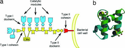
Fig. 1.
The cellulosome. (a) Schematic of the cellulosome. The type I dockerins, appended to the catalytic subunits, interact with the cohesin modules on the scaffoldin (CipA) leading to the formation of the supramolecular cellulosome complex. The type II dockerin on CipA, by binding to a type II cohesin on the bacterial membrane, tethers the cellulosome to the surface of C. thermocellum. (b) Internal symmetry of the WT dockerin in complex with cohesin. Not only do residues 1–22 overlap with 35–56, but the reverse is also true, because the dockerin shows internal 2-fold symmetry (panel b adapted from ref. 18).

