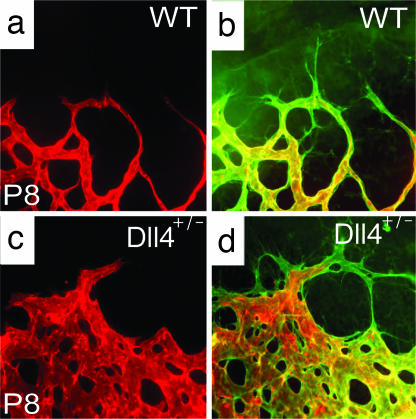
Fig. 3.
Perfusion staining of developing retinal vasculature in Dll4 mice. (a–d) Perfusion staining with L. esculentum lectin (a and c) and counterstaining with GS lectin (green, b and d) of the developing retinal vasculature in wild-type (a and b) and Dll4+/lacZ (c and d) mice at P8. (Original magnification: ×630.)

