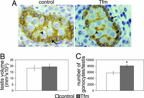
Fig. 1.
Morphometric analysis of 17.5 dpc fetal testes. (A) Histologic appearance of the testis in control and testicular feminized (Tfm) fetuses. Sertoli cells (arrows) were immunostained with anti-mullerian hormone and located at the periphery of the seminiferous cord, whereas gonocytes (arrowheads) were the large unstained cells in the center of the cord. (Scale bars, 10 μm.) (B and C) Testicular volume (B) and number of gonocytes per testis (C) in Tfm mice and their normal littermates. All results are shown as the mean ± SEM; n = 5. ∗, P < 0.05 in Student's t test.

